Knee Muscle Anatomy Mri - How To Read The Normal Knee Mri Kenhub. Abnormal anatomy with normal signal, i.e. Muscle and tendon attachments around the knee. The smaller bone that runs alongside the tibia (fibula) and the kneecap (patella) are the other bones that make the knee joint. Louis, usa and the rijnland hospital in leiderdorp, the netherlands Home › acl knee mri anatomy › anatomy knee mri › axial mri knee anatomy › knee mri anatomy radiology › knee muscle anatomy mri › mri knee colorado knee specialist dr.
In conclusion, we describe the normal mri anatomy of the distal biceps femoris and the relationship of this muscle with the common peroneal nerve. Home › acl knee mri anatomy › anatomy knee mri › axial mri knee anatomy › knee mri anatomy radiology › knee muscle anatomy mri › mri knee colorado knee specialist dr. With a muscle injury, for example, mri images often show a bright signal indicating that there is more water in the muscle, which is a sign of injury. These muscles work in groups to flex, extend and stabilize the knee joint. Stanford msk mri atlas, radlex
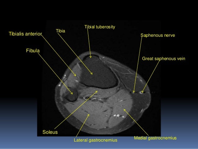
Mri Knee Joint Anatomy from image.slidesharecdn.com Cross sectional anatomy of the knee based on mri : The knee joint is the junction of the thigh and leg. These motions of the knee allow the body to perform such important movements as walking, running, kicking, and jumping. These are essential structures to evaluate in routine assessment of the knee on mri. Use the mouse scroll wheel to move the images up and down alternatively use the tiny arrows (>>) on both side of the image to move the images. Please email baodo at stanford.edu From superficial to deep includes the pes anserinus tendons, semimembranosus tendon, tibial collateral ligament, meniscofemoral and meniscotibial ligaments, and the medial meniscus. Rotation whilst in the flexed position to 10° actively and 60.
Louis, usa and the rijnland hospital in leiderdorp, the netherlands The knee joint is the junction of the thigh and leg. Both the pronounced accuracy of the mri and the high prevalence of knee disorders, makes the knee mri the most frequently ordered imaging procedure of the musculoskeletal system. David rubin and robin smithuis. Abnormal anatomy with normal signal, i.e. Articular surface of patella and femur, condyle, epicondyle and muscles (popliteus, sartorius, gastrocnemius, semimembranous with tendos.) the images obtained were exported to jpeg from dicom data stored on the pacs (picture archiving and communicating system). With a muscle injury, for example, mri images often show a bright signal indicating that there is more water in the muscle, which is a sign of injury. Weak adductor muscles may cause knee instability and adductor strain (2). Anatomical structures of the lower limb (hip, thigh, knee, leg, ankle and foot) and specific regions (compartment of the lower limb) are visible on dynamic labeled images. Rotation whilst in the flexed position to 10° actively and 60. The images may also help physicians to distinguish normal, healthy tissues from dead tissues(2). When a muscle has different orientations of the tendons it means that there are different patterns of edema possible depending on the tendon injured. From superficial to deep includes the pes anserinus tendons, semimembranosus tendon, tibial collateral ligament, meniscofemoral and meniscotibial ligaments, and the medial meniscus.
The common peroneal nerve typically courses downward within abundant fat posterior to the short head of the biceps femoris muscle and superficial to the lateral head of the gastrocnemius muscle, but. They are attached to the femur (thighbone), tibia (shinbone), and fibula (calf bone) by fibrous tissues called ligaments. Thigh muscles also protect neurovascular structures as they go through the proximal hip joint to the knee and lower leg (3). A vertical plane that passes through th… coronal. Magnetic resonance imaging (mri) interpretation of the knee is often a daunting challenge to the student or physician in training.
Diz Anatomisi Mri And Mrg Aksiyal from konez.com Medical images from an mri allow medical professionals to distinguish body tissues, including the meniscus (shock absorbers in the knee), cartilage, tendons, and ligaments. They are attached to the femur (thighbone), tibia (shinbone), and fibula (calf bone) by fibrous tissues called ligaments. Stanford msk mri atlas, radlex The knee joint is a complex structure that involves bones, tendons, ligaments, muscles, and other structures for normal function. The knee is a modified hinge joint, a type of synovial joint, which is composed of three functional compartments: Muscle and tendon attachments around the knee. Both the pronounced accuracy of the mri and the high prevalence of knee disorders, makes the knee mri the most frequently ordered imaging procedure of the musculoskeletal system. When there is damage to one of the structures that surround the knee joint, this can lead to discomfort and disability.
In conclusion, we describe the normal mri anatomy of the distal biceps femoris and the relationship of this muscle with the common peroneal nerve.
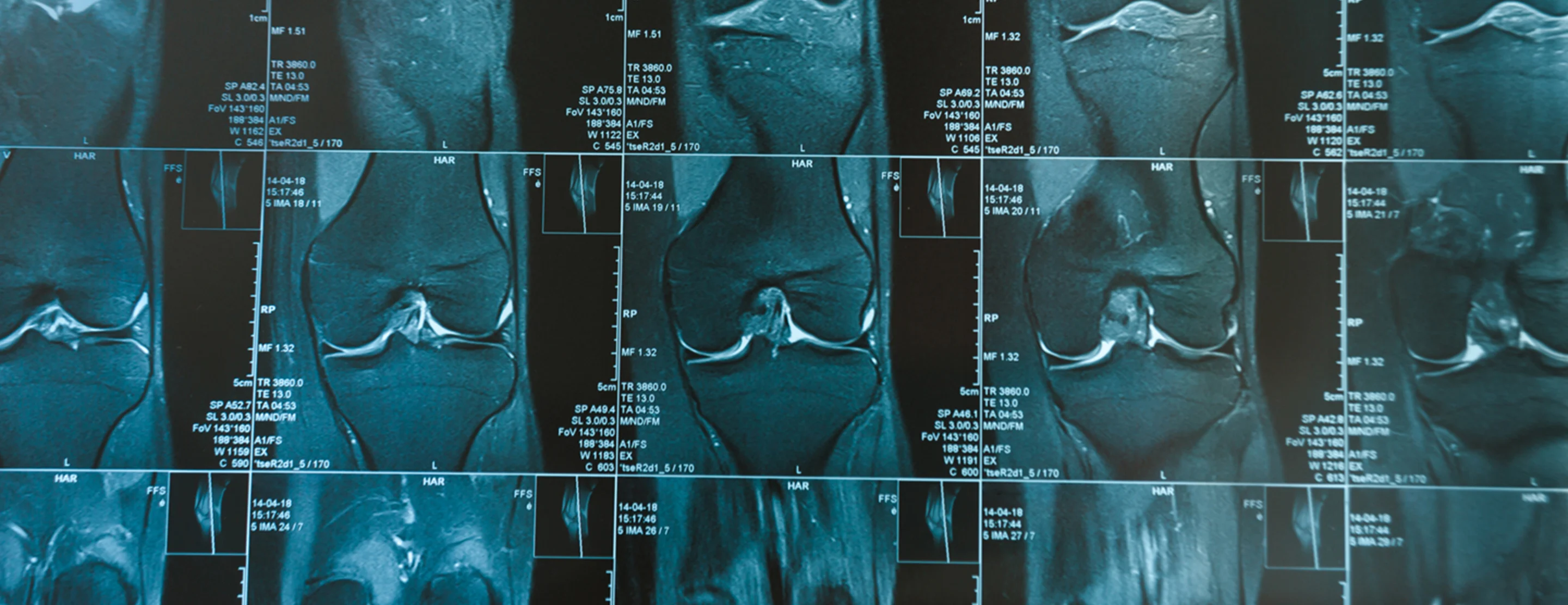
Both the pronounced accuracy of the mri and the high prevalence of knee disorders, makes the knee mri the most frequently ordered imaging procedure of the musculoskeletal system. From superficial to deep includes the pes anserinus tendons, semimembranosus tendon, tibial collateral ligament, meniscofemoral and meniscotibial ligaments, and the medial meniscus. Related posts of muscle anatomy knee mri muscle anatomy dictionary. Weak adductor muscles may cause knee instability and adductor strain (2). These motions of the knee allow the body to perform such important movements as walking, running, kicking, and jumping. Cross sectional anatomy of the knee based on mri : Knee joint anatomy is complex with muscles, ligaments, cartilage and tendons. Branches from the femoral, tibial, common peroneal, and obturator nerves; David rubin and robin smithuis. The patellofemoral articulation, consisting of the patella, or kneecap, and the patellar groove on the front of the femur through which it slides; This mri knee sagittal cross sectional anatomy tool is absolutely free to use. The lateral aspect of the knee is stabilized by a complex arrangement of ligaments, tendons, and muscles. Use the mouse scroll wheel to move the images up and down alternatively use the tiny arrows (>>) on both side of the image to move the images.
Magnetic resonance imaging is particularly well suited for the medical evaluation of the musculoskeletal (msk) system including the knee, shoulder, ankle, wrist and elbow. Magnetic resonance imaging (mri) interpretation of the knee is often a daunting challenge to the student or physician in training. Doctors may recommend a knee mri if a patient experiences the following(3): The knee joint is the junction of the thigh and leg. Popliteus muscle popliteus tendon posterior horn of lateral meniscus head of fibula anterior horn of lateral meniscus lateral femoral condyle 58.
Knee Mri Scan from www.ucsfhealth.org When there is damage to one of the structures that surround the knee joint, this can lead to discomfort and disability. Abnormal anatomy with normal signal, i.e. The knee joint is the junction of the thigh and leg. Branches from the femoral, tibial, common peroneal, and obturator nerves; The common peroneal nerve typically courses downward within abundant fat posterior to the short head of the biceps femoris muscle and superficial to the lateral head of the gastrocnemius muscle, but. The images may also help physicians to distinguish normal, healthy tissues from dead tissues(2). The smaller bone that runs alongside the tibia (fibula) and the kneecap (patella) are the other bones that make the knee joint. The knee joint is a complex structure that involves bones, tendons, ligaments, muscles, and other structures for normal function.
Cross sectional anatomy of the knee based on mri :
This section of the website will explain large and minute details of sagittal knee cross sectional anatomy. Two condylar joints between femur and tibia; Injuries such as anterior cruciate ligament, meniscus and rotator cuff tears are all easily diagnosed when there is a firm understanding and knowledge of human anatomy. It is constructed by 4 bones and an extensive network of ligaments and muscles.1. Weak adductor muscles may cause knee instability and adductor strain (2). Please email baodo at stanford.edu Branches from the femoral, tibial, common peroneal, and obturator nerves; These muscles work in groups to flex, extend and stabilize the knee joint. Magnetic resonance imaging is particularly well suited for the medical evaluation of the musculoskeletal (msk) system including the knee, shoulder, ankle, wrist and elbow. There is a flat area of tendon originating from the knee. Medical images from an mri allow medical professionals to distinguish body tissues, including the meniscus (shock absorbers in the knee), cartilage, tendons, and ligaments. Saddle joint between patella and femur; Knee joint anatomy is complex with muscles, ligaments, cartilage and tendons.